Home

Aktentasche Kalender schwindlig lvot echo Wange Beschwerde Fitness

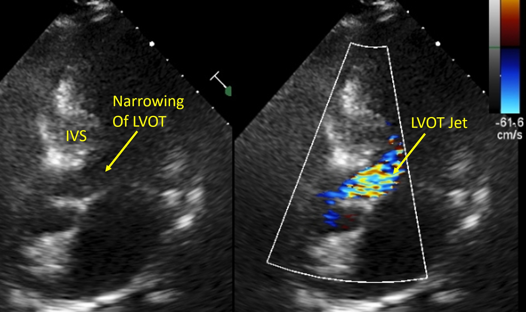
Routine orthostatic LVOT gradient assessment in patients with basal septal  hypertrophy and LVOT flow acceleration at rest: please stand up in: Echo  Research and Practice Volume 6 Issue 1 (2019)
Routine orthostatic LVOT gradient assessment in patients with basal septal hypertrophy and LVOT flow acceleration at rest: please stand up in: Echo Research and Practice Volume 6 Issue 1 (2019)
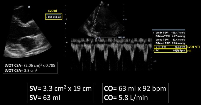
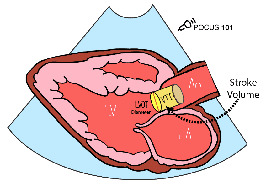
Rationale for using the velocity–time integral and the minute distance for  assessing the stroke volume and cardiac output in point-of-care settings |  The Ultrasound Journal | Full Text
Rationale for using the velocity–time integral and the minute distance for assessing the stroke volume and cardiac output in point-of-care settings | The Ultrasound Journal | Full Text

Normal Blood Flow Patterns

Left Ventricular Outflow Tract Obstruction in Hypertrophic Cardiomyopathy  Patients Without Severe Septal Hypertrophy | Circulation: Cardiovascular  Imaging
Left Ventricular Outflow Tract Obstruction in Hypertrophic Cardiomyopathy Patients Without Severe Septal Hypertrophy | Circulation: Cardiovascular Imaging

Cardiac Output by Echo - INTENSIVE

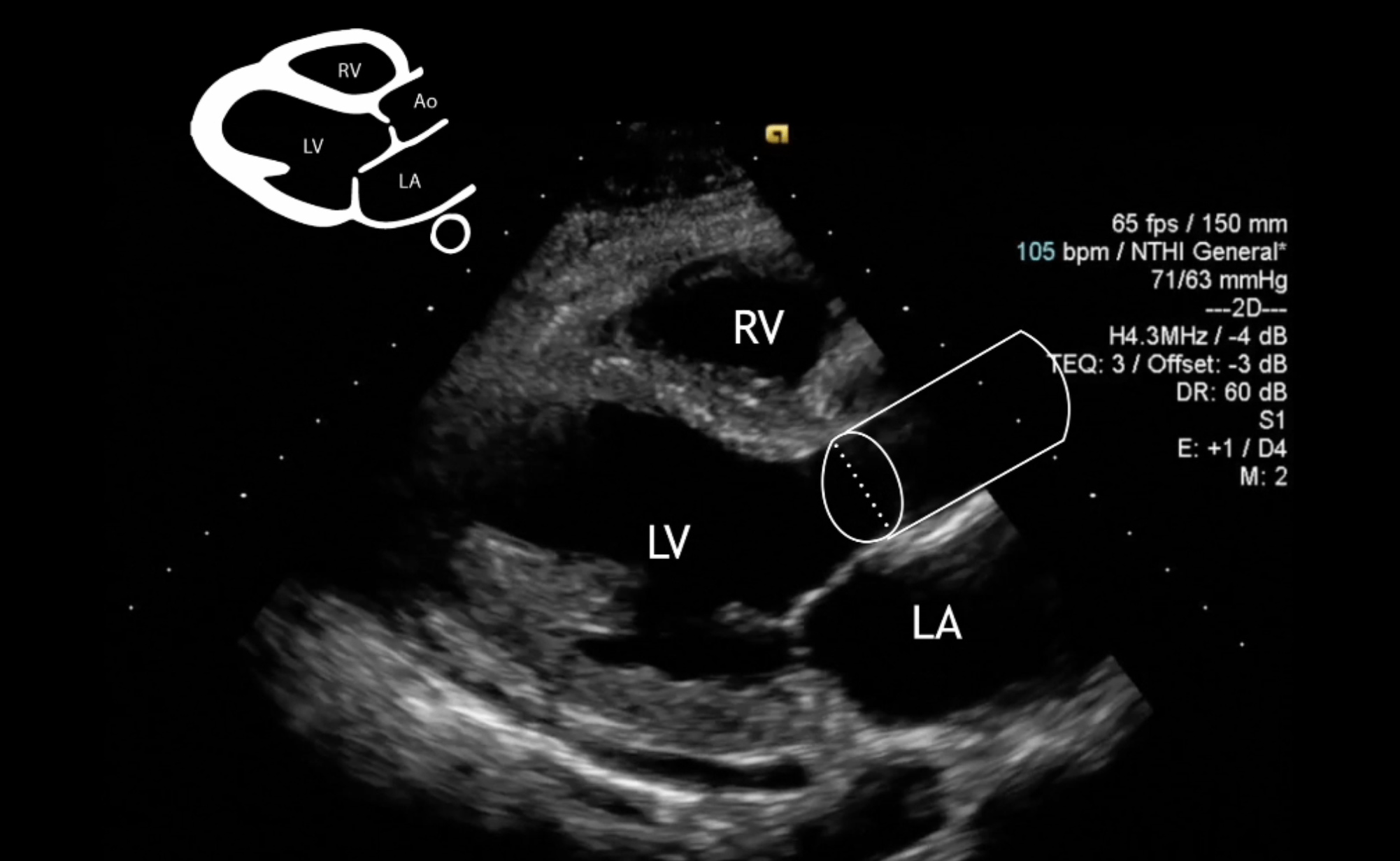
Measuring Cardiac Output with Echocardiography Made Easy - POCUS 101

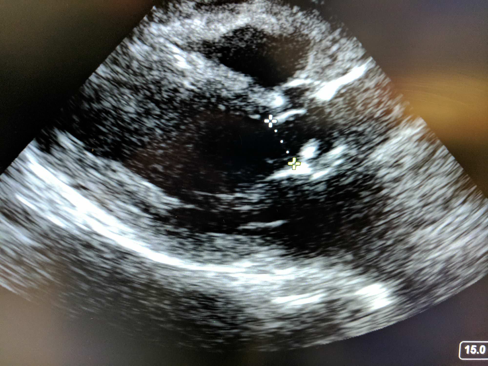
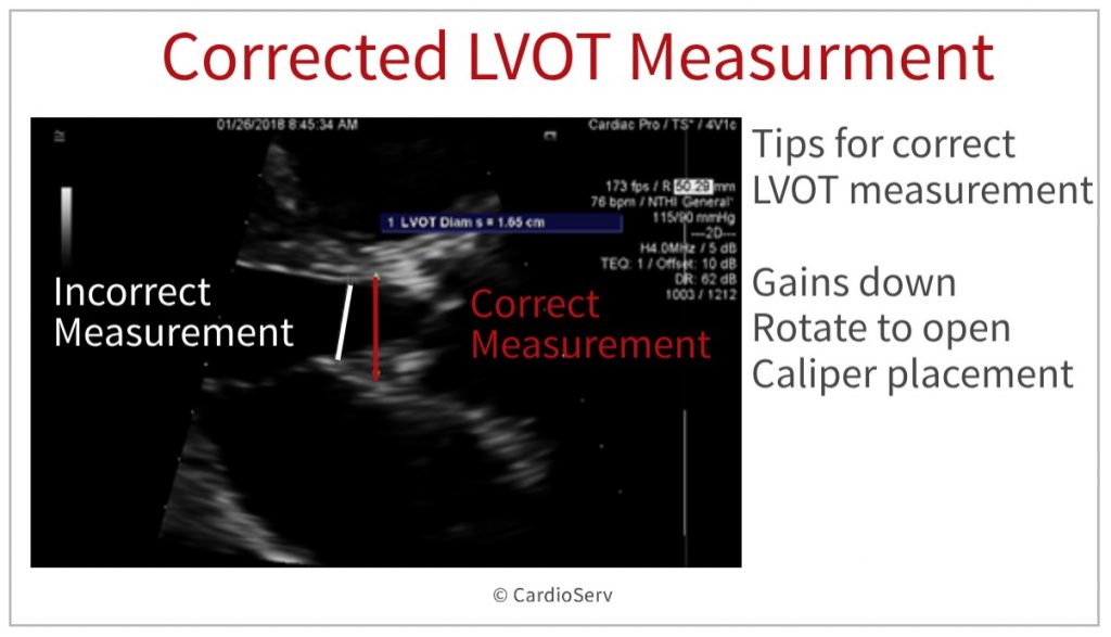
Accurate Measurement of Left Ventricular Outflow Tract Diameter: Comment on  the Updated Recommendations for the Echocardiographic Assessment of Aortic  Valve Stenosis - Journal of the American Society of Echocardiography
Accurate Measurement of Left Ventricular Outflow Tract Diameter: Comment on the Updated Recommendations for the Echocardiographic Assessment of Aortic Valve Stenosis - Journal of the American Society of Echocardiography

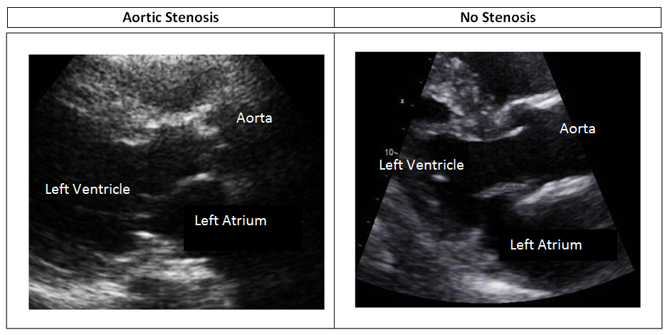
Aortic Stenosis: Breaking Down the Continuity Equation

Impact of anatomical variations of the left ventricular outflow tract on  stroke volume calculation by Doppler echocardiography in aortic stenosis -  Pu - 2020 - Echocardiography - Wiley Online Library
Impact of anatomical variations of the left ventricular outflow tract on stroke volume calculation by Doppler echocardiography in aortic stenosis - Pu - 2020 - Echocardiography - Wiley Online Library

IMPACT OF 3D ECHO ON THE ASSESSMENT OF AORTIC STENOSIS SEVERITY

Left ventricular outflow tract obstruction in echocardiography  (differential) | Radiology Reference Article | Radiopaedia.org
Left ventricular outflow tract obstruction in echocardiography (differential) | Radiology Reference Article | Radiopaedia.org
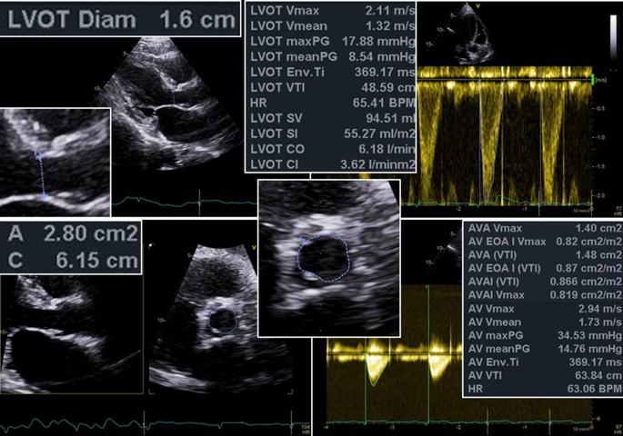
Cardiac Output With LVOT Measurements Using Echocardiography - RK.md

ECHOCARDIOGRAPHY: How to measure the LVOT? - YouTube

How to Master Aortic Measurements With These 5 Techniques

Expert consensus document on the assessment of the severity of aortic valve  stenosis by echocardiography to provide diagnostic conclusiveness by  standardized verifiable documentation | SpringerLink
Expert consensus document on the assessment of the severity of aortic valve stenosis by echocardiography to provide diagnostic conclusiveness by standardized verifiable documentation | SpringerLink

A) Long-axis TOE view showing the left ventricular outflow tract... |  Download Scientific Diagram
A) Long-axis TOE view showing the left ventricular outflow tract... | Download Scientific Diagram

Direct Planimetry of Left Ventricular Outflow Tract Area by Simultaneous  Biplane Imaging: Challenging the Need for a Circular Assumption of the Left  Ventricular Outflow Tract in the Assessment of Aortic Stenosis -
Direct Planimetry of Left Ventricular Outflow Tract Area by Simultaneous Biplane Imaging: Challenging the Need for a Circular Assumption of the Left Ventricular Outflow Tract in the Assessment of Aortic Stenosis -

Normal canine echo - right parasternal long axis LVOT view - YouTube

Disclosures
Disclosures

Aortic Stenosis and Mismatch Values

Accurate stroke volume (SV) estimation: SV = LVOT area × LVOT VTI. a... |  Download Scientific Diagram
Accurate stroke volume (SV) estimation: SV = LVOT area × LVOT VTI. a... | Download Scientific Diagram

PW Doppler LV outflow - UpToDate

Aortic Valve Area Calculation in Aortic Stenosis by CT and Doppler  Echocardiography - ScienceDirect
Aortic Valve Area Calculation in Aortic Stenosis by CT and Doppler Echocardiography - ScienceDirect

Accessory mitral valve leaflet causing left ventricular outflow tract  obstruction in an adult | Heart
Accessory mitral valve leaflet causing left ventricular outflow tract obstruction in an adult | Heart

Cardiac Output
Cardiac Output

Echo quiz – All About Cardiovascular System and Disorders

Transthoracic echo is used to assess AS severity and aortic annular... |  Download Scientific Diagram
Transthoracic echo is used to assess AS severity and aortic annular... | Download Scientific Diagram